
Once the diagnosis has been made, we need to know how far the cancer extends in the organ where it is located and whether it has spread to other organs (metastasis). This means “staging” the tumor by means of instrumental examinations: CT, PET, magnetic resonance, scintigraphy, echography, etc.
Depending on the stage of the tumor, the first important decision regarding treatment will be taken. The options are:
- operate immediately (or administer local therapies instead of operating);
- administer treatment first, and then operate;
- administer treatment without operating, as surgery is no longer possible because the disease is already at an advanced stage.
The first step in staging is to find out whether there are any distant metastases (WHAT ARE METASTASES?).
Metastases are groups of cells that have detached from the tumor and spread to other organs, where they have begun to grow and to form other nodules.
The question of whether or not there are distant metastases is of enormous importance (WHY STAGING IS SO IMPORTANT). If there are already metastases, the disease is generally no longer curable; if, by contrast, the tumor has not yet given rise to metastases, the disease can generally be cured by removing the tumor surgically.
With regard to distant metastases, staging can have 3 possible outcomes (FIGURES 2 and 3):
- positive for the presence of metastases;
- negative, because there are no metastases;
- negative, because metastases are present but are still too small to be seen (less than 0.5 – 1 cm). In this last case, the disease is actually at an advanced stage, though neither the patient nor the doctor can know this.
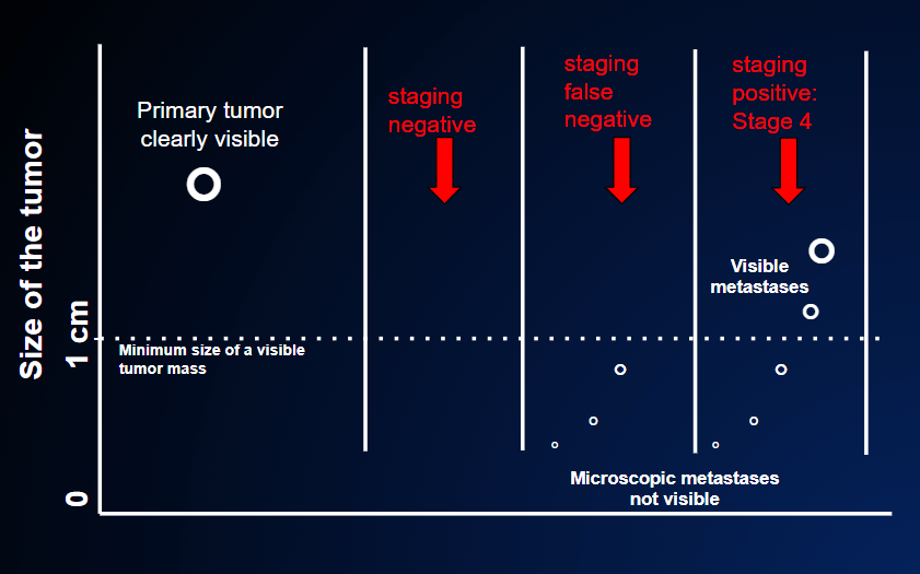
In the second case, the staging result is falsely negative because although the CT, PET scan and magnetic resonance tests are negative (=no tumor in other organs) the primary tumor has already spread outside the local origin, but these small nodules are too small to be revealed by the imaging tests. These tests usually disclose nodules of at least 0.5-1 cm ( the dotted line in the figure). After the primary tumor is removed, the patient is not cured. A relapse will become known when these small nodules have grown above that threshold.
In the third case, the staging result is positive (= presence of cancer in other organs ) because metastases are already seen by the staging tests: they are already large enough at the time the primary cancer is discovered.
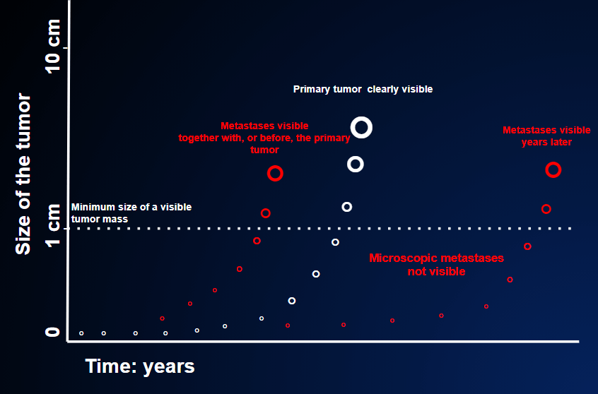
There is, however, an important difference between the first and the third conditions described above. In general, the first situation is one of disseminated (widespread) disease that is no longer curable, while in the third situation, cure is still possible. Indeed, postsurgical therapy – so-called adjuvant therapy – is able to eliminate these tiny (less than 1 mm) deposits of tumor cells (micrometastases), precisely because they are microscopic. In the first situation (metastases of a few cm), by contrast, it is not. (POSSIBLE PREVENTIVE THERAPY AFTER SURGERY)
The second step in staging, when distant metastases have been ruled out, is to establish the local extension of the tumor, in order to determine how easy or difficult it will be to completely remove it by means of surgery.
Local staging depends on the size and local extension of the tumor and on whether the nearby lymph nodes are affected.
The table below shows the classic division of tumors into 4 stages. The differences are extremely important, as they enable us to clearly distinguish between conditions that are curable by means of surgery and other therapies (stages 1, 2 and 3) and those which are generally no longer curable owing to the advanced stage of the disease (stage 4).
CLASSIC STAGING: 4 LEVELS OR STAGES
| STAGE | TUMOR | LOCAL LYMPH NODES | METASTASES |
|---|---|---|---|
| 1 | small | without tumor | absent |
| 2 | larger | without tumor | absent |
| 3 | large or small | with tumor | absent |
| 4 | large or small | with or without tumor | present |
In reality, however, things are much more complicated than this, as each type of tumor has its own variants. Nevertheless, this simple classification provides a general indication of the gravity of tumors.
Finally, it must be pointed out that the preoperative staging described above will be completed when surgery is performed. Indeed, when the surgeon directly inspects the operating field and the entire surgical specimen is analyzed by the pathologist, the stage previously assigned to the tumor on the basis of imaging may be confirmed or modified.